Indice del volumen Volume index
Comité Editorial Editorial Board
Comité Científico Scientific Committee
ONE-AND-A-HALF SYNDROME. CLINICAL CLASSIFICATION.
Foyaca-Sibat H, Ibanez-Valdes LdeF.
University of Transkei.Faculty of Health Sciences. Department of Neurology. Umtata, South Africa.
Rev Electron Biomed / Electron J Biomed 2004;3:15-21.
Comment of the reviewer Dr. R. Díaz-Alersi Hospital Puerto Real. Cádiz. Espańa
Comment of the reviewer Dr. JM. Trejo Gabriel y Galán. Hospital General Yagüe. Burgos. Espańa
Comment of the reviewer Dr. M. Fernández Albán. Centro de Investigaciones Médico Quirúrgicas (CIMEQ), La Habana, Cuba.
Key Words: One-and-a half syndrome, internuclear ophthalmoplegia. Neurocysticercosis (NCC), brainstem lesions
ABSTRACT
We studied 19 patients fulfilling clinical diagnostic criteria of One-and-a-half syndrome (OAHS) and all of them were grouped in three different types according to their neuro-ophthalmological disturbances. A novel clinical classification is suggested. Patients presenting OAHS secondary to racemose neurocysticercosis were reported for the first time to the medical literature.
RESUMEN:
Sindrome del Uno y Medio. Clasificación clínica.
Se estudiaron 19 pacientes que presentaron clinicamente un "Síndrome del Uno y Medio" en diferentes variantes y de causas diversas. Se agruparon los pacientes en 3 grupos diferentes de acuerdo a sus características neuro-oftalmológicas evitando ańadir nuevos eponimos a la ya existente lista de trastornos de la motilidad ocular. Proponemos una clasificacion clínica no
reportada con anterioridad y se reporta como causa novedosa la forma
racemosa de la neurocisticercosis para una de las variantes clínicas.
INTRODUCTION
The One-and-a half syndrome (OAHS) is a neurological disorder of the extra ocular movements characterized by conjugate horizontal gaze palsy (CHGP) in one direction and internuclear ophthalmoplegia (INO) in the other. The commonest cause of the syndrome is a vascular lesion on the basilar territory followed by demyelinating lesions of the brainstem usually affecting the paramedian pontine reticular formation (PPRF) unilaterally or due to lesions on the abducens nucleus (AN) on one side, with interruption of internuclear fibers of the ipsilateral medial longitudinal fasciculus (MLF) after it has crossed the midline from its side of origin in the contra lateral abducens nucleus (causing failure of adduction of the ipsilateral eye)1
OAHS type II has been described in patients presenting loss of voluntary eyes movements except for adduction in one eye usually secondary to lesions on the cerebral hemisphere and the cavernous sinus2 or unilateral infarction of the midbrain and thalamus3 the main difference between type I and type II consisted in preservation of abduction in one eye (type I) or sparing of adduction for one eye (type II), other combinations of paralysis of the eyes movements have been reported to the medical literature 4-8 but one attempt to classify this disorder in a single syndrome has not been made. The aim of this study is to report our clinical and aetiological findings in patients presenting OAHS of several aetiologies, and other associations. A novel clinical classification for this syndrome is suggested.
MATERIAL AND METHODS
We studied 19 consecutive patients admitted at stroke unit and neurological wards from Umtata General Hospital (UGH) and Nelson Mandela Academic Hospital (NMAH), between January 1998 and January 2004. All patients presented diverse modalities of horizontal/vertical gaze paralysis to one side and contra lateral jerking nystagmus or abduction/adduction paralysis on the opposite eye. Exclusion criteria were: Upper motor or sensory signs on the limbs, altered level of consciousness, signs of peripheral neuropathy, signs of cognitive dysfunction, pregnant patients and patients without written consent.
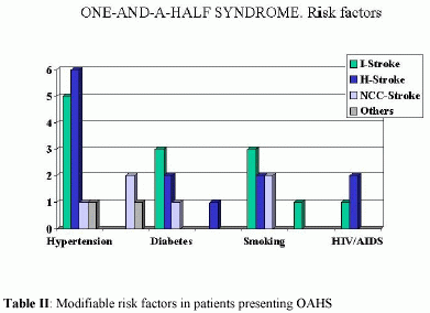
A senior neurologist (FSH) examined all patients in this series. We analysed the modifiable risk factors for stroke including cigarette smoking, hypertension, diabetes mellitus, hyperlipidemia, hyperuricemia, venous hematocrit (>50), coagulation screen, electrolytes, migraine, heart disease, arrhythmias, peripheral vascular diseases, and CT brain scan of patients from both hospitals and for patients from NMAC we also analysed protein S, protein C, antithrombin III, other causes of thrombophilia, and CD4 count-viral load when necessary. Investigations for multiple sclerosis were not done because that disease does not exist in this region.
All patients were distributed in three groups named type I (t-I), type II (t-II) and type III (t-III) according with their neuro-ophthalmologic disorders. Patients presenting CHGP and internuclear ophthalmoplegia or CHGP and preserved abduction of one eye were selected for t-I; patients presenting CHGP and sparing adduction of one eye or papillary disturbances were grouped in t-II, and others with CHGP plus unilateral vertical paresis or other combinations were selected for t-III group. Clinical and etiological correlation was done.
RESULTS
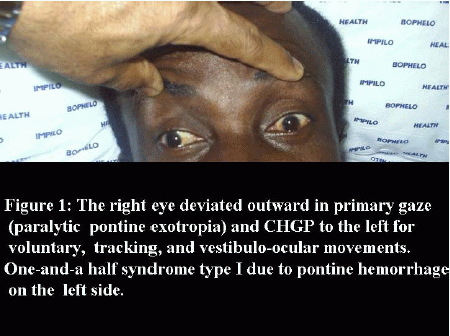
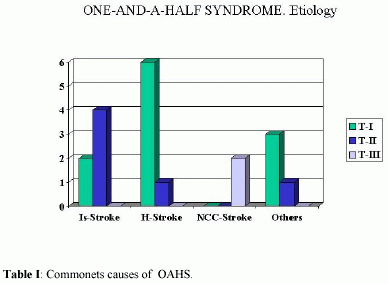
Nine patients presented with clinical features of t-I (47.3%), t-II characteristic were confirmed in 5(26.3%), and 2 were classified as t-III (10.5%). The commonest presentation of t-I was CHGP to the left and paralytic pontine exotropia of the right eye (Figure 1) and the more frequent cause of this syndrome was hemorrhagic stroke (Table I).
Patients with chronic and uncontrolled arterial hypertension were more commonly observed in our series (table II).
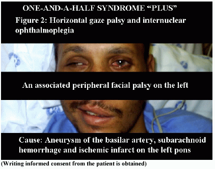
One patient with OAHS t-I also presented an associated peripheral facial palsy, and ipsilateral dilation of the pupil secondary to subarahcnoid hemorrhage, paramedian tegmental infarct, and aneurysmatic dilatation of the basilar artery HIV/AIDS related (Figure 2).
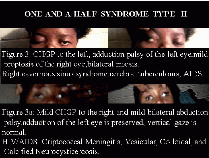
Ischemic stroke was the aetiology of OAHS t-II in 10.5% of this series and cavernous sinus syndrome and midbrain infarction were present in only one patient being the most rare cause of this syndrome (Table I) Cavernous sinus syndrome and an associated proptosis of the affected side was also seen (Figure 3) however typical CHGP and preserved adduction eye movement due to hemorrhagic stroke was seen in only one patient.
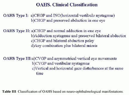
One patient presented conjugate vertical gaze palsy (CVGP) and limited downward movements in one eye and other presented combined CHGP and limitation on the vertical movements in one eye. All patients with limitations for eyes movements in our series were grouped in 3 types as can be see on Table III
DISCUSSION:
Type I
It is a clear-cut, well-known and worldwide accepted syndrome and the correlation between CHGP and INO or adduction on one eye is almost pathonogmonic of pontine lesion due to stroke or multiple sclerosis affecting the AN, PPRF, and ipsilateral MLF. Several reports to the medical literature have been made9-13 If there is damage of the vestibular fibres from horizontal semicircular canal to the medial vestibular nucleus (MVN) or from the MVN to the AN then horizontal vestibular nystagmus is present, this happens because MVN send excitatory impulse to the contralateral AN and inhibitory impulses to the ipsilateral one and because there is saccadic input to the abducens from the ipsilateral excitatory PPRF and contralateral inhibitory input from the contralateral and dysfunctional state of this mechanism can cause nystagmus. Pathogenesis for exotropia of one eye has been clearly described by Jhkura et al.14 See Table III
Type II:
As before mentioned this variant was reported previously by Carter and Rauch2 in 1994 and two years later by Celibisoy and Akyureki,3 their reports were not congruent from the clinical and pathological point of view but their description has been positively accepted, Nevertheless, one patient of the 19 patients with OAHS type II reported by Thomke15 presented an associated vertical gaze paresis of unknown cause, we consider that patients complaining of vertical eyes involvement should not be included in this group because obviously the mechanism for this disorder differ from the one accepted for this variant. Our clinical findings were also different as well.
However, we accept as valid this type of OAHS for patients presenting CHGP and preservation of adduction movements on one eye during saccadic and pursuit eye movements or an associated papillary abnormalities if there is no disturbances of the vertical gaze and if more than one lesion of the CNS can be demonstrated. See Table III
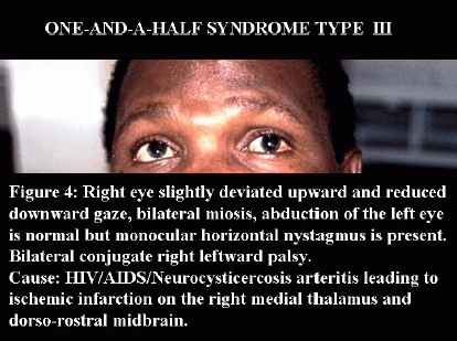
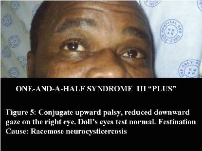
Type III
Bogousslavsky and Regli16 reported one case of vertical "One-and-a-half syndrome (VOAHS) twenty years ago, their patient had an upgaze palsy and monocular paresis of downward secondary due to an infarct of the paramedian thalamus and upper mesencephalom on the right side affecting the fibres of the posterior commisure and descending fibres to the ipsilateral subnucleus of the inferior rectus and sparing fibres of the subnucleus of the superior oblique just after they decussate. From the same authors similar reports but due to different causes years later were made17, 18.
In 1989, Deleu et al19reported one patient with VOAHS characterized by supranuclear downgaze paralysis with monocular elevation palsy secondary to bilateral infarction in the mesodiencephalic region affecting the efferent tracts of the rostral interstitial nucleus of the MLF bilaterally and the premotor fibres to the contralateral superior rectus subnucleus and ipsilateral inferior oblique subnucleus, either before or after decussating in the posterior commissure, since then combinations of CVGP downward or upward and upward or downward monocular paresis has been nominated as VOAHS however combinations of CHGP and CVGP remained out of classification up to date. Cases reported by Terao et al20 and our patients fulfilling criteria to be grouped in the same category and other reports before cited also coincide with similar aetiological lesions at the mesencephalo-diencephalic level therefore there is no reason to exclude those patients from this syndrome.
Our patients presenting CVGP and monocular vertical disturbances or CVGP and CHGP and all had more that one ischemic vascular lesion at the midbrain and thalamic-mesencephalic level probable secondary to infectious vasculitis HIV-NCC related. This association of racemose NCC and OAHS is being reported to the medical literature for the first time.
In 1998, Eric Eggenberg9 reported three cases of isolated OAHS with facial palsy secondary to ischemic infarction on the vertebrobasilar territory, he added 7 (Cranial nerve number 7) to 1 and ½ (OAHS) and he termed that combination as Eight-and-half syndrome, we found patients with other combinations that can be nominated as Five-and-a-half syndrome (1 ½ + 4-throclear nerve), Seven-and-a-half syndrome (1 ½ + 6-abducens) or Eleven-and-a-half syndrome (1 ½ + 7-facial nerve + 3-oculomotor nerve) however we honestly believe that generation of novel arithmetic combination will bring more confusion than clarity to this problem and its will increase unnecessarily the long list of different names of neuro-ophthalmological disorders, perhaps adding the term "plus" will be enough to distinguish this condition from the typical OAHS t-I. We prefer and recommend the term of OAHS for all presentations and the sub-term of "type I-II-III" to separate one combination from another, this classification will be useful for an accurate identification of its aetiologies also. If patients present an associated motor or sensory signs then they should no be included in this classification and a diagnosis of Locked-in syndrome, vertebro-basilar stroke or other disorder should be done accordingly.
Acknowledgments: We thank to Prof. Awotedu, Prof. Targoska and Dr. Anwary for their support.
REFERENCES:
-
1. - Wall M, Wray SH. The one-and-a half syndrome a unilateral disorder of the pontine tegmentum: a study of 20 cases and review of the literature. Neurology 1983;33(8):971-980
2. - Carter JE, Rauch RA. The one-and-a half syndrome, type II. Arch Neurol 1994;51:373-377.
3. - Celibisoy N, Akurekli O. One-and-a half syndrome, type II: a case with rostral brainstem infarction. Neuro-ophthalmology 1996;16:373-377
4. - Deleu D, Buisseret T, Ebinger G. Vertical one-a-and syndrome. Supranuclear downgaze paralysis with monocular elevation palsy. Arch Neurol 1989;46(12):
5. - Kataoka S, Hori A, Shirakawa T, Hirose G. Paramedian Pontine Infarction. Neurological/Topographical Correlation Stroke 1997;28:809-815
6. - Terao S, Osano Y, Fukuoka T, Miura N, Mitsuma T. Coexisting vertical and horizontal one and a half syndromes J Neurol Neurosurg Psychiatry 2000;69:401-402
7. - Bhidayasiri E et al Ahypothetical scheme for the brainstem control of vertical gaze. Neurology 2000;54:1985-1993
8. - Onofrj T et al Clinically evidenced unilateral dissociation of saccades and pursuit eye movements. J.Neurol Neurosurg Psychiatry 2004;75:1084-1050
9. - Wall M, Wray SH, The one-and-a-half syndrome a unilateral disorder of the pons tegmentum: a study of 20 cases and review of the literature. Neurology 1983;33(8):971-980
10. - Martin PJ, Chang HM, Wityk R, Caplan LR. Midbrain infarction: associations and aetiologies in the New England Medical Center Posterior Circulation Registry. J Neurol Neurosurg Psychiatry 1998;64:392-395
11. - De Seze J, Lucas C, Leclerc X, Sahli A, Vermersch P, Leys D. One-and-a-half syndrome in pontine infarcts: MRO correlates. Neuroradiology 1999;41(9):666-669
12. - Minagar A, Schatz NJ, Glaser JS. Case Report: One-and-a-Half Syndrome and Tuberculosis of the Pons in a Patient with AIDS. AIDS PATIENT CARE and STDs 2000:14(9):461-464
13. - Kumral E, Gamse B, Akyol A, Yunten N, Sirin H, Sagduyu Y. Mesencephalic and Associated Posterior Circulation Infarcts. Stroke 2002;33:2224-2236
14. - Jhkura K, Komiyama A, Kuroiwa Y. Eye deviation in patients with One-and-a-Half Syndrome. Eur Neurol 2000;44:210-215
15. - Thomke F. The so-called one-and-a half syndrome type II: a new syndrome? Neuro-ophthalmology 1999;22(2):73-79
16. - Bogousslavky J, Regli F. Upgaze palsy and monocular paresis of downward gaze from ipsilateral thalamo-messencephalic infarction: vertical "one-and-a-half" syndrome. J Neurol 1984;231(1):43-45
17. - Bogousslavki J, Meisnberg O. Eye-movement disorders in brain-stem and cerebellar stroke. Arch Neurol 1978;44:141-148
18. - Hommel M, Bogousslavki J. The spectrum of vertical gaze palsy following unilateral brainstem stroke. Neurology 1991;41:1229-1234
19. - Deleu D, Buisseret T, Ebinger G. Vertical one-and-a-half syndrome. Arch Neurol 1989;46(12):1361-1363
20. -Eggenberger E. Eight-and-a-half syndrome plus cranial VII palsy. J.Neuroophthalmol 1998;18:114-116
Comment of the reviewer R. Díaz-Alersi MD. Hospital Puerto Real. Cádiz. Espańa
Advanced neuroimaging techniques has been replacing the topographic diagnosis usually made by clinical examination.
Nevertheless, there are moments in which an accurate clinical examination done by skilled hands can substitute neuro-imaging techniques including MRI for topographic diagnosis specially in case where those procedures are contraindicated. One-and-a-half syndrome is a good example of the before cited. It is a uncommon disease described for the first time by Fisher in 1997 and is characterized by horizontal conjugate gaze palsy and an associated ipsilateral internuclear ophthalmoplegia in other word; complete paralysis of horizontal eyes movement in one side and impaired adduction of the contralateral eye.
This syndrome is usually due to a pontine lesion affecting the parapontine reticular formation nucleus, abducens nucleus, middle longitudinal fascicules, lateral vestibular nucleus and its efferent pathways. The commonest cause is a vascular lesion on the basilar territory or demyelinating disorders. The biggest series of patients reported to the medical literature is made by Wall et al, they presented 49 patients.
Foyaca-Sibat et al presented a series of 19 cases fulfilling the diagnosis criteria for One-and-a-half syndrome that were studied between 1998 and 2004. Author suggested a novel clinical classification for this syndrome and reported the first patient presenting this syndrome due to neurocysticercosis.